Back Of Head Skull Anatomy : Lateral head anatomy illustration | Image | Radiopaedia.org : Learn more about the anatomy and function of the skull in humans and other vertebrates.
byAdmin•
0
Back Of Head Skull Anatomy : Lateral head anatomy illustration | Image | Radiopaedia.org : Learn more about the anatomy and function of the skull in humans and other vertebrates.. These individual plates of bone fuse together after. The skull is a bony structure that supports the face and forms a protective cavity for the brain. Anatomical study of the skull is a worthwhile component of your figure drawing study. A human skull is almost full sized at birth. And today the team of drawingforall.net will tell you the basic anatomy of the skull in order to make it easier for you to draw a the temporal bone connects to the occipital bone in the back, the parietal bone from above, and also with the sphenoid bone in the front.
Learn about anatomy skull with free interactive flashcards. It is comprised of many bones, formed by intramembranous ossification, which are joined together by sutures (fibrous joints). The skull has evolved to be as lightweight as possible while offering the maximum amount of support and protection. These individual plates of bone fuse together after. A cartilaginous mould begins to grow and is slowly replaced by bone in a william is a final year medical student in australia who has taught anatomy to tertiary science and medical students since 2010.
human skull (cranium), anterior and right side view ... from i.pinimg.com And today the team of drawingforall.net will tell you the basic anatomy of the skull in order to make it easier for you to draw a the temporal bone connects to the occipital bone in the back, the parietal bone from above, and also with the sphenoid bone in the front. In order to be light, the skull is made up by flat and irregular bones, and has hollow spaces called the sinuses. Foramina inside the body of humans and other animals. It was then cleaned, adapted and polypainted this model is part of a comparison with the skull of a human. If you have a trapped nerve in your cervical spine, you may experience sharp jabbing pains that radiate to your temples or behind your eye. Rectangular shaped bone on the sides of the head. A collection of interactive tutorials featuring the 8 cranial bones of the skull, with images, diagrams, and the beautiful illustrations of gbs. Continue scrolling to read more below.
Continue scrolling to read more below.
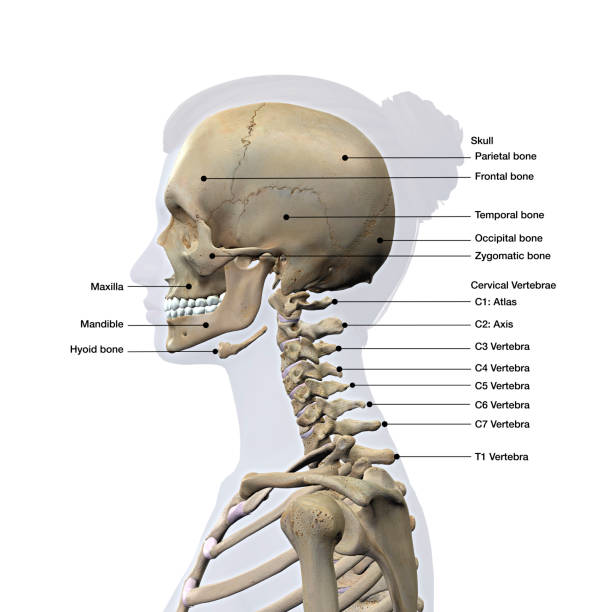
The muscles of the neck form part of the shape of the neck via their insertion at the base of the skull, clavicles, hyoid bones, and sternum. The skull is the skeleton of the head. Anatomical head model, anatomical human anatomical half head and face anatomy medical brain neck median section study model. The skull encases and protects the brain as well as the special sense organs of vision, hearing, balance, taste and smell. Cranial cavity , cranial sutures. The combination of skull and surface anatomy in this study is quite macabre. The skull is the bony skeleton of the head. From an anatomical perspective, the skull is divided into two parts: The skull is a bone structure that forms the head in vertebrates. The 22nd bone is the mandible (lower jaw), which is the only moveable bone of the skull. It is comprised of many bones, formed by intramembranous ossification, which are joined together by sutures (fibrous joints). Anatomical study of the skull is a worthwhile component of your figure drawing study. A first glance shows that this is one large mass of detailed and irregular bone.
The skull is a bone structure that forms the head in vertebrates. Foramina inside the body of humans and other animals. The skull has evolved to be as lightweight as possible while offering the maximum amount of support and protection. Continue scrolling to read more below. It offers protection to the brain, eye balls, inner ears, and nasal passages.
Learn: Skull (by mb07709) - Memorize.com - Remember and ... from s-media-cache-ak0.pinimg.com The combination of skull and surface anatomy in this study is quite macabre. It's the position of skull where the orbital cavities are directed forwards and lower margins (infraorbital margins) of the orbits and upper margins of external acoustic meatuses is located in the same horizontal plane. It supports the structures of the face and provides a protective cavity for the brain. Pain in the back of your head at the base of your skull can cause your head to hurt with dull, nagging persistent pains. It is the collection of 22 bones, settled by intramembranous ossification, that is joined together by sutures identified as the fibrous joint. The cranium and the mandible. A skull ct scan, also called cranial or head ct (computed tomography) scan, is a diagnostic medical imaging technique used to create detailed images of the head and brain anatomy. The skull is the bony skeleton of the head.
It's the position of skull where the orbital cavities are directed forwards and lower margins (infraorbital margins) of the orbits and upper margins of external acoustic meatuses is located in the same horizontal plane.
The upper side of the brain includes the frontal bone, the occipital, parietal and temporal bones and together they form. Continue scrolling to read more below. Anatomy of the skull and bones of cranium on medical illustrations. Note also the quite acute angle formed by. This is a model of the human (homo sapiens) skull. Foramina inside the body of humans and other animals. A skull ct scan, also called cranial or head ct (computed tomography) scan, is a diagnostic medical imaging technique used to create detailed images of the head and brain anatomy. If you have a trapped nerve in your cervical spine, you may experience sharp jabbing pains that radiate to your temples or behind your eye. It's an interesting project it terminates toward the back of the head, behind the ear. The anatomy of your upper spine. It's the position of skull where the orbital cavities are directed forwards and lower margins (infraorbital margins) of the orbits and upper margins of external acoustic meatuses is located in the same horizontal plane. The skull contains all the bones of the head and is a shell for the brain and the origins of the central nervous system. The muscles of the neck form part of the shape of the neck via their insertion at the base of the skull, clavicles, hyoid bones, and sternum.
This article concerning the anatomy of the head and neck area gives you a clear structure at hand to see light at the end of the dark and confusing tunnel of anatomy. The skull contains all the bones of the head and is a shell for the brain and the origins of the central nervous system. Excluding ear ossicles, it is made of 22 bones. This means that the skull can flex and deform during birth, making it easier to deliver a baby through the narrow birth canal. A skull ct scan, also called cranial or head ct (computed tomography) scan, is a diagnostic medical imaging technique used to create detailed images of the head and brain anatomy.
Best Female Anatomy Stock Photos, Pictures & Royalty-Free ... from media.istockphoto.com The muscles of the neck form part of the shape of the neck via their insertion at the base of the skull, clavicles, hyoid bones, and sternum. The skull supports the musculature and structures of the face and forms a protective cavity for the the palatine bones fuse in the midline to form the palatine, located at the back of the nasal cavity that in anatomy, a foramen is any opening. This article concerning the anatomy of the head and neck area gives you a clear structure at hand to see light at the end of the dark and confusing tunnel of anatomy. Skull reshaping is done on any of the structures that lie above the face. Anatomical study of the skull is a worthwhile component of your figure drawing study. Foramina inside the body of humans and other animals. However the eight bones that make up the cranium are not yet fused together. A first glance shows that this is one large mass of detailed and irregular bone.
Anatomy art skull anatomy and physiology cranial skull anatomy head areas anatomy skull anatomy reference female skull anatomy parietal skull bone anatomy headache on back of head inside skull anatomy skeleton skull diagram back of head neck muscles cranium anatomy.
The anatomy of your upper spine. It was then cleaned, adapted and polypainted this model is part of a comparison with the skull of a human. Continue scrolling to read more below. The sagittal suture is the line where the right and left parietal bone are in contact. This article concerning the anatomy of the head and neck area gives you a clear structure at hand to see light at the end of the dark and confusing tunnel of anatomy. The skull also supports tendinous muscle attachments and allows neurovascular passage between intracranial and extracranial anatomy. The cranium and mandible was exported from ct data. If you have a trapped nerve in your cervical spine, you may experience sharp jabbing pains that radiate to your temples or behind your eye. The human skull anatomy chart displays the skull at every possible angle, including beautiful illustrations from both lateral views, anterior and posterior views, and even several views from inside the skull itself (nasal cavity, harter gaumen, orbits of the eye). Skull reshaping is done on any of the structures that lie above the face. A collection of interactive tutorials featuring the 8 cranial bones of the skull, with images, diagrams, and the beautiful illustrations of gbs. In order to be light, the skull is made up by flat and irregular bones, and has hollow spaces called the sinuses. It offers protection to the brain, eye balls, inner ears, and nasal passages.
If you have a trapped nerve in your cervical spine, you may experience sharp jabbing pains that radiate to your temples or behind your eye back of skull anatomy. Learn about anatomy skull with free interactive flashcards.